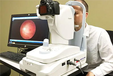
Fundus photography involves capturing a photograph of the back of the eye. Specialized fundus cameras that consist of an intricate microscope attached to a flash enabled camera are used in fundus photography.
Anterior segment eye photography is rapidly becoming a valuable part of vision care. A simple 35 mm SLR camera set-up that can be easily operated without an extensive photographic background has been designed from readily available equipment.
Eyeglasses lenses correct refractive errors by focusing light directly on the retina. The type of lens depends on the type and severity of the refractive error. First we do the diagnosis using the finest machines and find the errors like Myopia, Hyperopia, Presbyopia and Astigmatism.
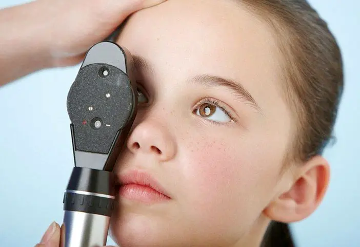
Direct ophthalmoscopy one that produces an upright, or unreversed, image of approximately 15 times magnification.
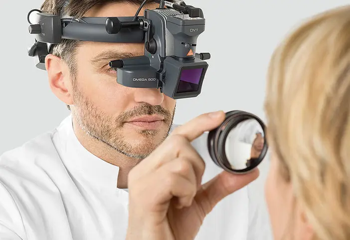
The indirect ophthalmoscope is mounted on the head of the viewer and a condensing lens is held close to the eye. The viewer is about arms-length away from the patient. It provides the viewer a much wider field of vision of the back of the eye and the view shows elevation and depth like 3D. Almost all eye examinations by ophthalmologists today utilize the indirect ophthalmoscope.
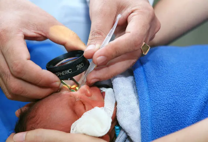
ROP is a pathologic process that occurs only in immature retinal tissue and can progress to a tractional retinal detachment, which can result in functional or complete blindness.

The +90-D lens exhibited a linear relationship between p and degree of ametropia of the model eye and a constant relationship between p and ametropia of −5 to +5 D. As far as we are aware, this has not been reported previously for the +90-D lens.
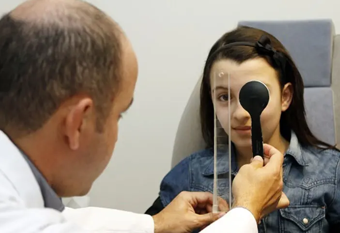
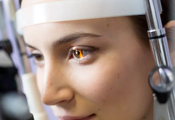
The examiner uses a retinoscope to shine light into the patient's eye and observes the reflection (reflex) off the patient's retina. While moving the streak or spot of light across the pupil the examiner observes the relative movement of the reflex or manually places lenses over the eye (using a trial frame and trial lenses) to "neutralize" the reflex.
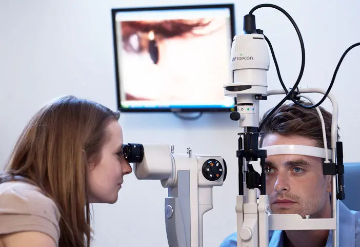
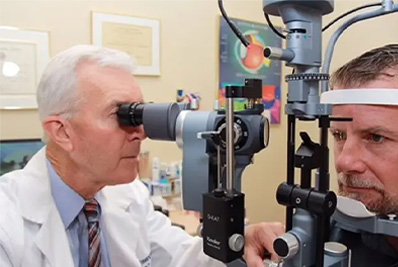
The slit lamp is an instrument consisting of a high-intensity light source that can be focused to shine a thin sheet of light into the eye.